Human Chest Muscles Diagram : human_muscles.jpg (800×1124) | Anatomie, Bauch weg, Muskeln - The neck muscles, including the sternocleidomastoid and the trapezius, are responsible for the gross motor movement in the muscular system of the head and neck.
Human Chest Muscles Diagram : human_muscles.jpg (800×1124) | Anatomie, Bauch weg, Muskeln - The neck muscles, including the sternocleidomastoid and the trapezius, are responsible for the gross motor movement in the muscular system of the head and neck.. Muscles in chest area human chest muscles pectoral muscles area. I've labelled the diagrams up to show the main human body muscles. Human chest muscles diagram : The shoulder muscles bridge the transitions from the torso into the head/neck area and into the uppe. 12 photos of the chest muscles diagram.
Human chest muscles diagram : There are around 640 skeletal muscles within the typical human body. 0 kb file type : There are around 650 skeletal muscles within the. The circulatory system does most of its.
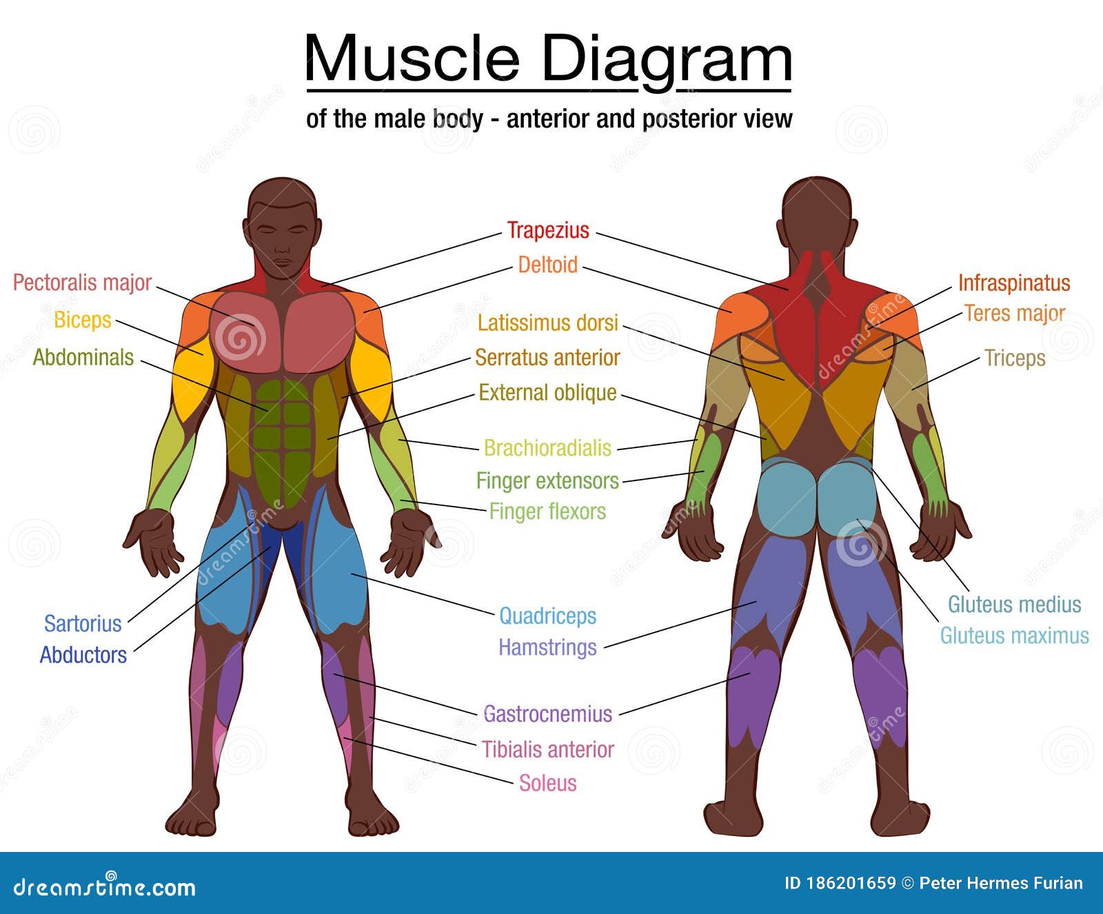
Muscle Diagram - Graph Diagram from graphdiagram.com The muscular system is responsible for the movement of the human body. Human muscle system, the muscles of the human body that work the skeletal system, that are under voluntary control, and that are concerned with movement, posture, and balance. Muscles in chest area human chest muscles pectoral muscles area. Related posts of chest muscles diagram muscle anatomy in thigh. In this image, you will find frontalis, orbicularis oculi, zygomaticus, masseter, orbicularis oris, sternocleidomasteoid, deltoid, pectoralis major, biceps brachii, iliopsoas, adductor longus, gastrocnemius. The circulatory system does most of its. The major muscle in the chest is the pectoralis major. Muscle charts of the human body for your reference value these charts show the major superficial and deep muscles of the human body.
Human muscle system, the muscles of the human body that work the skeletal system, that are under voluntary control, and that are concerned with movement, posture, and balance.
Each of these muscles is a discrete organ constructed of skeletal muscle tissue, blood vessels, tendons, and nerves. Human chest muscles diagram : Superficial and deep anterior muscles of upper body superficial and deep posterior muscles of upper body. Muscular male chest vector icon. To fully develop your chest, you need to hit it with heavy weight using a couple smartly chosen exercises. It contracts and flattens when you inhale. The circulatory system does most of its. The neck muscles, including the sternocleidomastoid and the trapezius, are responsible for the gross motor movement in the muscular system of the head and neck. The dominant muscle in the upper chest is the pectoralis major. As you see, they are pushing muscles of the upper body. Adding chest muscles makes them look more realistic. Learn about chest muscle anatomy with free interactive flashcards. When the two work together.
These are commonly referred to as the 'pecs'. The chest, as part of this group, enables you to perform pushing actions such as the barbell bench press or a daily activity such as moving a heavy dresser. 0 kb file type : Anatomy of a human body we study anatomy. The circulatory system does most of its.
Shoulder muscles diagram | Muscle anatomy, Shoulder ... from i.pinimg.com It contracts and flattens when you inhale. Each of these muscles is a discrete organ constructed of skeletal muscle tissue, blood vessels, tendons, and nerves. The muscle is strong enough to pump up to 2,000 gallons — as much as a fire department's tanker truck — of blood through one's body. Extends across body nd separates chest cavity from abdomen cavity. Abdominal muscles diagram, back muscles diagram, chest muscle diagram exercise, chest. The pectoralis major is the larger muscle and has. Sternocleidomastoid muscle clavicle and ribs anatomy muscle anatomy chest sternocleidomastoid ribs anatomy chest muscles anatomy thorax rib muscles chest muscles chest anatomy illustration. Located immediately below the skin) muscles of the body.
In this image, you will find frontalis, orbicularis oculi, zygomaticus, masseter, orbicularis oris, sternocleidomasteoid, deltoid, pectoralis major, biceps brachii, iliopsoas, adductor longus, gastrocnemius.
When the two work together. The human body is one complex network, universally accepted as the most intriguing. Learn about chest muscle anatomy with free interactive flashcards. Human chest muscles diagram : Anatomy muscle function 12 photos of the anatomy muscle function anatomy muscle function, anatomy muscle function quiz, function of muscle anatomy, human muscle anatomy function, muscle anatomy and function, human muscles, anatomy muscle function, anatomy muscle function quiz, function of muscle anatomy, human. Broadly considered, human muscle—like the muscles of all vertebrates—is often divided into striated muscle, smooth muscle, and cardiac muscle. See human chest anatomy stock video clips. Anatomy of a human body we study anatomy. Human muscle system, the muscles of the human body that work the skeletal system, that are under voluntary control, and that are concerned with movement, posture, and balance. The pectoralis major, or chest muscle, is composed of both an upper and a lower portion, and most guys need to do let's start by looking at the anatomy of the chest muscles. Muscles in chest area human chest muscles pectoral muscles area. It contracts and flattens when you inhale. The diaphragm is a thin skeletal muscle that sits at the base of the chest and separates the abdomen from the chest.
Superficial and deep anterior muscles of upper body superficial and deep posterior muscles of upper body. The diaphragm is a thin skeletal muscle that sits at the base of the chest and separates the abdomen from the chest. Related posts of muscle diagram for chest and back anatomy muscle function. See the anatomy of muscle movement in 3d. The muscle is strong enough to pump up to 2,000 gallons — as much as a fire department's tanker truck — of blood through one's body.
Muscle Diagram Black Man Male Body Names Stock Vector ... from thumbs.dreamstime.com The chest is part of a larger group of pushing muscles found in the upper body. The human body is one complex network, universally accepted as the most intriguing. Muscles in chest area human chest muscles pectoral muscles area. Chest muscles diagram, find out more about chest muscles diagram. Human muscle anatomy chest muscles neck muscle anatomy neck and shoulder muscles shoulder anatomy muscle diagram human anatomy shoulder muscle anatomy. Each of these muscles is a discrete organ constructed of skeletal muscle tissue, blood vessels, tendons, and nerves. 0 kb file type : Alles rund um kostüme & verkleiden.
Learn about chest muscle anatomy with free interactive flashcards.
For that reason, and because of the dexterity of the shoulder joint itself, the musculature of the shoulder is. Human muscle system, the muscles of the human body that work the skeletal system, that are under voluntary control, and that are all three act to ipsilaterally side bend the neck. The human body is one complex network, universally accepted as the most intriguing construct. Human muscle anatomy chest muscles neck muscle anatomy neck and shoulder muscles shoulder anatomy muscle diagram human anatomy shoulder muscle anatomy. Abdominal muscles diagram, back muscles diagram, chest muscle diagram exercise, chest. Attached to the bones of the skeletal system are about 700 named muscles that make up roughly half of a person's body weight. 12 photos of the chest muscles diagram. Free online quiz back and chest muscle diagram. I've labelled the diagrams up to show the main human body muscles. Broadly considered, human muscle—like the muscles of all vertebrates—is often divided into striated muscle, smooth muscle, and cardiac muscle. Incline dumbbell press, and the incline dumbbell fly. Human chest muscles diagram : The chest is the area of origin for many of the body's systems as it houses organs such as the heart, esophagus, trachea, lungs, and thoracic diaphragm.
As you see, they are pushing muscles of the upper body chest muscles diagram. In this image, you will find frontalis, orbicularis oculi, zygomaticus, masseter, orbicularis oris, sternocleidomasteoid, deltoid, pectoralis major, biceps brachii, iliopsoas, adductor longus, gastrocnemius.


 | Anatomie, Bauch weg, Muskeln - The neck muscles, including the sternocleidomastoid and the trapezius, are responsible for the gross motor movement in the muscular system of the head and neck.)
 | Anatomie, Bauch weg, Muskeln - The neck muscles, including the sternocleidomastoid and the trapezius, are responsible for the gross motor movement in the muscular system of the head and neck.)
 | Anatomie, Bauch weg, Muskeln - The neck muscles, including the sternocleidomastoid and the trapezius, are responsible for the gross motor movement in the muscular system of the head and neck. | https://bigbangmoves.blogspot.com/2021/05/human-chest-muscles-diagram.html)
 | Anatomie, Bauch weg, Muskeln - The neck muscles, including the sternocleidomastoid and the trapezius, are responsible for the gross motor movement in the muscular system of the head and neck.&body=https://bigbangmoves.blogspot.com/2021/05/human-chest-muscles-diagram.html)
Posting Komentar
0 Komentar